What is a ureteral stone and why is it dangerous?
Ureteral stone is an independent nosological form of urolithiasis caused by obstruction of the lumen of the upper urinary tract. People of different age categories are more or less at risk of developing this pathology. Pediatric urolithiasis is detected in boys and girls with almost the same frequency. Among adult patients, men predominate (3:1). In this case, the main age difference lies in the structure of urolith stones.
The main danger of all stones, regardless of biomineralogical composition, is their continuous growth. Wedged into the lumen of the ureter, the stone disrupts the outflow of urine, destabilizes the functioning of the entire excretory system, and causes painful attacks of renal colic. In 60% of cases, infectious inflammation occurs at the site of urolith localization. In the absence of timely treatment, the infection rises, affects the tubular system of the kidneys and provokes the development of acute pyelonephritis. If normal urine flow is not restored, pathogenic bacteria can enter the bloodstream and cause life-threatening urosepsis.
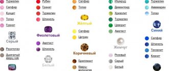
Varieties and colors
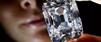
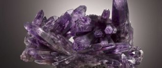
Red varieties of corundum are called rubies, all others (colorless, blue, violet, green, yellow, orange, pink) are called sapphires.
In general, the following varieties are distinguished for corundum as a mineral species:
· Ruby is a red stone of various shades; rubies of the “pigeon’s blood” color are especially in demand.
· Sapphires - come in blue, cornflower blue and colored (yellow, orange, pink, green, purple).
Leucosapphire is a colorless sapphire.
· Padparadscha is a pinkish-orange sapphire, although its color has a more poetic definition: the color of “a lotus flower in the rays of the setting sun in Sri Lanka.”
· Star ruby and star sapphire are gemstones with the effect of asterism, which is manifested by observing the shape of a six- or twelve-pointed star when light hits a cabochon-shaped surface.
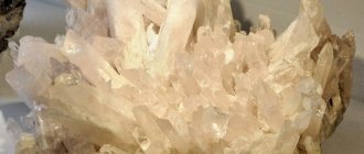
· Ordinary corundum is an opaque coarse- or fine-grained stone of a grayish color.
· Emery is a granular corundum rock of gray-black color.
· Synthetic (artificial) corundum - lilac-pink varieties are called “French rose” or “rose of France”.
The red color of ruby is due to an admixture of chromium, and the blue of sapphire is due to an admixture of titanium and iron. The yellow color is due to the presence of ferric iron.
Symptoms of a ureteral stone
Clinical manifestations depend on the location, degree of obstruction and rate of development of urinary tract obstruction. When a stone stops near the upper pole of the kidney, the main complaint is pain in the lower back. When the lower area is affected, another characteristic symptom of a stone in the ureter is added: in men - irradiation of pain into the testicle, in women - into the labia. If the stone moves downwards, the location of the colic may change.
Urination is accompanied by severe pain, and fresh blood appears in the urine. Ureteral pain spreads to the thoracic spine (region of 11-12 vertebrae). With complete obstruction, against the background of intense pain, nausea and vomiting develop, temperature indicators remain within normal limits.
Important!
An increase in body temperature to 38-39-40°C indicates the development of an infectious-inflammatory process. This is a very dangerous condition that requires immediate medical attention.
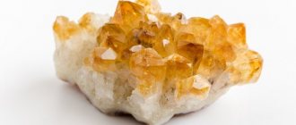
Opal - radiant sun
Opal received its second name - the stone of unfulfilled hopes. This mineral is perfect for Libra; it will be a real guardian and assistant for representatives of this air sign.
The stone radiates due to the presence of water in the composition. Opal is a precious stone and is often used in gold and silver jewelry. Previously, hairpins and hair decorations were often made for fashionistas using white opals. Young girls believed that precious stones could protect them from being deceived by the opposite sex during courtship.
Infusions with opal, like those with pearls, are also beneficial for the body. They cope well with gastrointestinal disorders and some infections of the genitourinary system. White opal protects against sore throat if you wear jewelry around your neck in the form of pendants or beads.
It is believed that opal received its name for a reason. A person who has lost hope experiences this very hard. White opal will help to cope with this; its reflections will calm the heart and soul, and will reveal to its owner faith in the best.
The gemstone is also suitable for Pisces and Scorpios. These zodiac signs rarely indulge in daydreaming, but sometimes it is necessary. For outwardly cold Pisces, opal will allow them to better express their emotions, and Scorpio may have a dream and a goal to realize it.
White opal protects its owner from tumor diseases. And if a malignant formation has already appeared, then the gemstone is able to restrain its growth. You can give such a pebble to a cancer patient. This is not a guarantee of a cure, but hope for the best. The mineral bestows faith in a positive future.
If the owner of the mineral has kidney disease, then wearing jewelry with opal is necessary; the stone has a beneficial effect on these organs and prepares the body to fight pathology.
If a person has experienced a great loss or is faced with grief, then the stone can restore interest in life and awaken a craving for change.
Precious opal also requires special care to prevent it from losing its shine or becoming brittle. It should be washed at least once a month under running water.
Causes of occurrence and development of urolithiasis
Stone formation is a pathological process associated with metabolic disorders. Urolithiasis is a multifactorial disease caused by various external and internal causes:
- geographical location and associated composition of drinking water;
- climatic conditions (lack of UV, vitamin D deficiency, chronic dehydration);
- pathologies associated with prolonged immobilization;
- harmful production factors;
- burdened heredity;
- developmental anomalies of the urinary system;
- increased secretion of parathyroid hormone, increased calcium concentration in the blood plasma;
- damage to bone tissue;
- kidney injuries;
- inflammatory urological diseases;
- pathologies leading to stagnation of urine and loss of urinary sediment;
- taking pharmaceutical drugs that crystallize and promote the formation of stones;
- consequences of radiation therapy.
The progressive development of urolithiasis occurs against the background of local changes in the renal pelvis. A prerequisite is an increased concentration of stone-forming ions, a lack of substances that slow down the crystallization process and the presence of stone growth activators in the urine.
Classification and stages of stone development in the ureter
All urinary stones known to science, in most cases, are formed in the renal pelvis, and enter the ureters along with the bloodstream. Based on the nature of their origin, they are divided into infectious (caused by a microbial pathogen) and non-infectious. The latter, in turn, are genetically determined (associated with hereditary disorders) and medicinal. Individual formations are formed from various mineral components, which over time consolidate into conglomerates.
According to the salt composition, ureteral stones are:
- urate (sodium, potassium salts of uric acid);
- phosphate (calcium hydrogen phosphate, struvite, carbonate apatite);
- oxalate (derivatives of oxalic acid);
- mixed (formed by various minerals).
Calculi of organic origin associated with disorders of amino acid metabolism include cystine, xanite and protein stones.
Urolithiasis has 4 stages of development:
I – (crystalluria): change in urine pH, increase in the concentration of organic substances, precipitation of salt crystals;
II – (microlithiasis): appearance of sand and small stones (< 4 mm)
III – formation of uroliths > 5 mm, disrupting the outflow of urine;
IV – obstruction of the ureter, impossibility of spontaneous passage of the stone, intractable colic.
What complications may arise?
In the absence of timely medical care, a stone in the ureter can cause:
- local inflammatory process;
- urethral syndrome (painful urination, the appearance of pus in the urine);
- pyelonephritis;
- ureteral or autonomic vascular spasm;
- disorders of blood circulation and concentrating ability of the kidneys;
- hydronephrosis;
- dysfunction of the excretory organ.
To prevent irreversible damage associated with unilateral or bilateral ureteral obstruction, it is necessary to seek medical help at the first symptoms of urolithiasis in men and women.
Diagnosis of ureteral stone
Urologists manage patients with signs of obstructive uropathy. Reasons to consult a doctor include pain, decreased daily urine output, or complete absence of urine. In a conversation with the patient, the existence of urolithiasis and other associated pathologies (neoplasms of the genitourinary organs, intestines, etc.) must be clarified.
Diagnosis of urolithiasis includes laboratory and instrumental (imaging) examination methods. To identify a stone in the ureter, the following is prescribed:
- general clinical urine test, blood biochemistry;
- Ultrasound of the excretory system: detection of stones in the kidneys, upper and lower ureteral segments;
- duplex scanning of renal vessels: assessment of ureteric output, renal blood flow, determination of the degree of obstruction;
- survey urography, X-ray diffraction study: differentiation of X-ray positive and X-ray negative stones;
- CT, non-contrast or with contrast (a more informative x-ray examination that allows you to determine the size, density, and exact location of the stone);
- MSCT;
- magnetic resonance urography: assessment of the current state of all soft tissue components of the collecting system.
Note!
The choice of the optimal method and sequence of instrumental diagnostics is carried out individually, taking into account the symptoms and the expected level of obstruction. Multislice computed tomography is recognized as the most informative and rational method for detecting ureteral stones.
What does a stone therapy procedure look like?
When a person first hears about stone therapy, most likely, he immediately remembers popular advertising photos of spa salons, which depict girls with polished black stones laid out in smooth lines on their bodies. But this is not the only technique used in stone therapy. Depending on the technique, the procedure can involve from 2 to 60 stones.
- Combined massage technique with hands and stones.
In this case, the stones are used as a tool for massage. There can be from 2 to 10. During the procedure, there may be no direct contact of the stone with the skin - in order to avoid burns [7]. If the client does not suffer from allergies, aromatic oils can also be used to enhance the beneficial effects of the massage.
- Massage exclusively with stones.
This type of stone therapy is a kind of symbiosis of acupressure and a heating pad. During the procedure, heated stones are applied to certain points on the body. For example, in case of cholelithiasis and cholecystitis, heat is applied to the spine. For angina pectoris, warm stones are placed on the left collarbone. To treat urinary incontinence, heat is placed on the upper sacrum. During such a procedure, the master can use about 60 stones. The classic set is 20 marble and 40 basalt.
The duration of one stone therapy session is about 60 minutes. But to achieve a visible cosmetic or therapeutic effect, it is recommended to undergo the procedure in courses.
Can a stone come out on its own?
This depends on the size of the stone, the individual characteristics of the structure of the excretory duct and the general condition of the patient. The normal width of the ureteral lumen is 5-7 mm. Small stones, not exceeding 4 mm, pass spontaneously in 80% of cases, without special medical intervention. 5 mm - in 50% of cases of the total.
Additional Information
Due to the fact that the junction of the renal pelvis and the ureter is irregular in shape and slightly narrowed in diameter, the risk of obstruction in this area increases significantly. True, such cases are quite rare in clinical practice. Most often, urinary stones are retained in the middle (45%) or lower (70%) part of the canal.
Treatment of ureteral stone
Treatment of urolithiasis includes therapeutic and surgical approaches. If the size of the stone does not exceed 7 mm, conservative correction is prescribed. Larger formations are removed promptly.
Conservative treatment
The complex of therapeutic measures includes:
- pharmacotherapy: antispasmodics, NSAIDs, corticosteroids, alpha-blockers, litholytic (stone-dissolving) agents, drugs to prevent relapses of urolithiasis, antibiotics (if a bacterial infection is attached);
- physiotherapy: exposure to laser, diathermic, amplipulse currents, vibration therapy, etc.;
- Exercise therapy: special sets of physical therapy exercises;
- diet therapy: nutritional correction, taking into account the nature of stone formation.
- sanatorium-resort treatment: drinking course of mineralized waters, therapeutic baths, physiotherapeutic procedures.
Surgery
Among the surgical techniques for treating urolithiasis in women and men, the following should be highlighted:
- remote shock wave lithotripsy (crushing of stones);
- endoscopic surgery (percutaneous, transurethral urethrolithotripsy);
- laparoscopic stone removal (urethrolithotomy).
Thanks to the active implementation of minimally invasive treatment methods and the multimodal strategy Fast Track surgery (postoperative accelerated rehabilitation), it has become possible to perform surgical interventions quickly, efficiently and effectively. Open abdominal surgery, due to its high morbidity and long recovery period, is currently used extremely rarely.
Urolithiasis disease
Urolithiasis (urolithiasis) in traditional official medicine is a disease associated with the formation of stones in the kidneys and/or other organs of the urinary system.
Urolithiasis can affect all age groups - from newborns to the elderly. The type of urinary stone usually depends on the age of the patient.
In older people, uric acid stones predominate. Protein stones form much less frequently. It should be noted that more than 60% of the stones are mixed in composition. Urinary stones almost always form in the kidneys. In most cases, they enter the ureter and bladder from the kidney. In most cases, KSD is a one-sided process, but sometimes stones are detected in both kidneys at once. The number of stones can vary widely - from single to multiple - several dozen. Stones can be small (2-3 mm) and large (up to 15 cm); observations of stones that weighed several kilograms have been described.
The main reason for the formation of kidney stones is metabolic disorders, especially changes in the water-salt and chemical composition of the blood. In addition to hereditary predisposition, risk factors for urolithiasis include dietary habits determined by the characteristics of national cuisine or the specific preferences of a particular patient.
The problem of so-called “secondary” stones stands apart - these are cases when stones are formed against the background of a violation of the outflow of urine and crystals of salts that are dissolved in high concentrations fall out in the form of sediment (crystallization theory of stone formation). The quality and chemical composition of drinking water is of great importance. Thus, there are well-known regions in which the incidence of urolithiasis is significantly higher than the national average. These include the Caucasus, the Volga region, and from foreign regions - Africa, the countries of Central and Southeast Asia, the islands of the Indian Ocean... Unfavorable factors for the development of ICD are: a sedentary lifestyle, a lack of vitamin A and B vitamins in food, the use of certain medications (sulfonamides, excessive consumption of ascorbic acid - vitamin C), as well as prolonged immobilization of the patient (consequences of injuries, fractures, etc.) Negative factors are chronic diseases of the gastrointestinal tract (gastritis, colitis, peptic ulcer, etc.) and organs genitourinary system (pyelonephritis, prostatitis, prostate adenoma, cystitis, etc.), dysfunction of the parathyroid glands; osteomyelitis, osteoporosis, other bone diseases or injuries; constant abuse of foods that increase the acidity of urine (spicy, sour, salty); drinking hard water with a high salt content;
Types of stones by composition.
Urates occur in 5–15% of people suffering from urolithiasis. These are stones consisting of uric acid and its salts (sodium and potassium). Urate stones are hard and smooth in structure, brick or yellow-orange in color. Due to their low density (the absence of calcium in their composition), urates are not visible on X-rays. They are diagnosed using ultrasound and laboratory urine analysis.
The reasons for the formation of such stones are poor nutrition, insufficient fluid intake (less than 2 liters per day), metabolic failure, and renal tubular disorders.
If you have urate stones, you should get tested for uric acid levels and rule out the development of a disease such as gout. Stones that arise due to the deposition of large amounts of uric acid salts can signal the development of joint diseases and vice versa.
Urate stones are the only ones that can be dissolved, especially if they are small in size. This requires alkalization of urine, a special diet, and the use of diuretics.
Oxalates are the most common type of stones. Formed in the kidneys due to an excess of calcium salts of oxalic acid. They have a high density, so they are easy to diagnose both with ultrasound and x-rays. Oxalates are high-density stones of black-gray color with a spiked surface. These spines often scratch the lining of the urinary tract, which can cause red blood cells to appear in the urine.
The movement of stones through the urinary tract can cause severe pain (renal colic). The pain can be localized in the lower back, groin area, and sides of the abdomen.
People who adhere to a healthy lifestyle often suffer from the formation of oxalate stones. These stones are formed by excessive consumption of citrus fruits and juices, sorrel, spinach, lettuce, beets, as well as tea, coffee and chocolate. Also, the risk of oxalate formation is high in people who consume small amounts of calcium, since this mineral binds and removes oxalic acid salts from the body. Other reasons for the formation of oxalate stones include vitamin B6 deficiency and certain diseases of the small intestine (resection, Crohn's disease).
Oxalate stones cannot be dissolved. If the stones are small (up to 4 mm), you can try to remove them from the body with urine. To do this, you need to drink a lot of fluid (up to 2.5 liters), stick to a diet and take measures to alkalize your urine. Passing a stone is a long and painful process, so you need to tune in to 3-4 weeks of treatment and, if necessary, relieve pain with antispasmodics and painkillers. If the stone is large, it must be removed. Most often, to remove oxalate stones, a technique such as lithotripsy is used - crushing stones using electromagnetic shock waves, puncture nephrolitholopaxy - crushing a stone after puncture of the kidney and inserting instruments into its cavity system, or contact lithotripsy - inserting instruments for crushing and extracting stone fragments through natural routes without additional incisions and punctures - through the urethra, bladder, ureter to the area where the stone is located. The extent of the operation depends on the location and size of the stone. Open surgery to remove the stone has become rare these days.
Phosphate stones are composed of calcium salts of phosphoric acid. Smooth or slightly rough stones have a soft consistency and white color. Most often they are formed in alkaline urine due to metabolic disorders. The occurrence of phosphates can be easily detected by doing a urine test - the pH value becomes greater than 6.2. If you see white, loose flakes in your urine, you most likely have phosphate stones.
Phosphates grow quickly, but nevertheless crush well. Treatment in this case should be aimed at acidifying the urine. This can be achieved by drinking sour juices, mineral waters, infusions of grape root, barberry, and rose hips. As a rule, as a result of such treatment, phosphate stones are easily crushed and at least stop increasing in size.
Struvite is a stone characterized by rapid growth and a soft structure. Their surface is smooth or rough, the color of such stones is white or light gray. Stones of this type are formed as a result of stagnation of urine or the activity of bacteria and are infectious in nature. Most often, struvite stones occur in women.
Struvite is dangerous because it can develop into coral stones. They are able to fill the entire kidney from the inside within a few months, creating an impression of the pelvis.
These stones are diagnosed using ultrasound, x-rays, computed tomography and urinalysis. If struvite stones form in the urine, under magnification, crystals similar in shape to the lid of a coffin are found.
In the case of the formation of struvite stones, treatment with herbal medicine and medications is ineffective. If the stone size is small, crushing on a lithotripter is used, and if the stone is large, surgery is needed. A situation often arises when getting rid of struvites using
If you experience renal colic, pain in the lower back, groin or sides, see your doctor immediately. Nephrolithiasis, detected in the early stages, is easily treatable and most often resolves without negative consequences.
Symptoms of urolithiasis
The classic manifestation of ICD is renal colic - an acute condition caused by a violation of the outflow of urine through the urinary tract. The nature and location of pain may depend on the position of the stone. Most often, stones in renal colic are detected in the area where the ureter originates from the pelvis or lower in the ureter. Until the moment when the stone manifests itself in this way, ICD may be asymptomatic. Renal colic is a sudden attack of severe pain in the lumbar region. Colic often occurs after a bumpy ride, heavy physical activity, or drinking large amounts of liquid. If the stone is located in the lower parts of the ureter, pain, in addition to the lumbar region, may appear in the lower abdomen and radiate to the groin area and external genitalia. The pain occurs suddenly at any time of the day. Changing body position does not affect the intensity of pain. Typical accompanying pain is nausea, vomiting and changes in the frequency of the urge to urinate, blood in the urine and pain when urinating. If the size of the stone does not exceed 5-6 mm (the diameter of the ureter), then it may pass on its own. Once in the bladder, the calculus most often comes out unhindered (the diameter of the urethra exceeds the diameter of the ureter). If the stone is large, remains in one place for a long time without a tendency to move, or is located in an area of narrowing of the urinary tract, specialist intervention may be required. Long-term disruption of the outflow of urine by a stone can cause inflammatory changes in the kidney or loss of its functional ability, resulting in shrinkage. After contacting a specialized institution, the minimum list of examinations includes blood and urine tests, ultrasound, and a plain X-ray (urography). A more in-depth study may include taking x-rays with preliminary introduction of radiopaque substances into the vein or computed tomography. An increase in body temperature to 38-40 degrees, which is typical for the addition of inflammation against the background of a violation of the outflow of urine.
A person can carry a kidney stone all his life and not know about it. But, on the other hand, a 3-4 mm stone that has begun its movement along the ureter can cause such renal colic that a person will literally climb the wall.
Treatment of urolithiasis.
First of all, when treating kidney stones, it is necessary to relieve an attack of renal colic. The next steps in treatment are removing the stone, treating the infection, and preventing the stones from reoccurring.
Currently, therapy for urolithiasis includes conservative and surgical treatment methods.
Conservative treatment - treatment by prescribing specific medications, following a diet and drinking regime. It can be quite effective if the kidney stones are small (up to 3-5 mm). If the inflammatory process begins, antibacterial therapy is also carried out. Systematic intake of antispasmodics and herbal diuretics is prescribed.
Diet for urolithiasis
Diet plays an extremely important role in the treatment of kidney stones. Its selection should be made by a doctor depending on the composition of the stones and what causes urolithiasis in a particular case. The diet limits or excludes from the patient’s diet foods that provoke the growth and formation of new deposits. The diet helps the stones pass and also helps soften them.
Diet for stones with a high urate content.
The goal of the diet for such stones is to reduce the level of uric acid and its salts in the body.
Number of meals per day: 5-6 with an equal break
What are the features of this diet:
ØFoods that are high in purine (a specific protein) are excluded from food.
Øat the same time, the normal content of proteins, fats, and carbohydrates in food is maintained
Øproducts that contain a sufficient amount of alkaline radicals are actively consumed (to increase the alkaline level)
What products are preferable: fermented milk products (including kefir and milk), various cereals (oats, buckwheat, millet), fruits (especially sweet ones), vegetables (almost any), natural juices (it’s better not to drink store-bought ones, as they may contain contain preservatives and taste regulators), animal proteins (eggs, lean fish, lean meat, chicken), for sweets it is better to use natural honey.
Foods to avoid: fried or smoked meat products, mushroom dishes, strong and caustic seasonings, products made from cocoa beans (including coffee, chocolate, cocoa), canned fish in any form.
It is better not to drink any alcohol during treatment.
Diet for phosphate stones.
The goal of the diet is to normalize the acid balance and thereby stop the appearance of calcium salts. This is achieved by increasing the content of vitamin A in food, increasing the acidity of urine, avoiding salt in food, water loading up to 2.5 liters of liquid per day, increasing the consumption of foods that contain calcium (including milk and dairy products, eggs, cheese).
Products that can be consumed for phosphaturia: low-fat fish and meat products (including boiled and even fried), various pasta, as well as soups (including with cereals and beans, only the broth for such a soup should not be too fatty), fruits (especially apples and currants, can be consumed in any form).
What is best to avoid with this diet: spicy foods, natural juices (from both fruits and vegetables and berries), dairy products are also not recommended for consumption, it is better to avoid them as much as possible. Avoid fruits and vegetables that are high in alkaline elements.
Alcohol will not benefit any diet. In addition, it is recommended to limit or minimize the consumption of drinks such as cocoa and coffee.
Diet for stones with a high content of oxalates.
The goal of the diet is to reduce the content of foods that contain oxalic acid. It is necessary to completely exclude from the diet such products as: sorrel, rhubarb, all cocoa derivatives (including chocolate). It is necessary to limit potatoes, carrots, tomatoes, onions, beets, and gelatin in the diet.
Foods that help lower oxalic acid levels: apples, grapes, plums and many other fruits.
During this diet, it is very important to drink plenty of fluids: about two liters, do not drink alcohol and reduce the amount of sweets (especially chocolate!). Fasting days will be very effective and useful, during which you are only allowed to drink juices, eat vegetables and apples (of course, those that are not prohibited for use in this diet).
Diet for high carbonate stones
You should carefully monitor the amount in food of those foods that increase the alkaline balance. Water load - at least two liters. The diet should not be too long - in itself it is not harmless to the body.
What foods should you limit?
First of all, these are any food products containing calcium, including: milk, yoghurts, cottage cheese, cheeses and other fermented milk products
beneficial for your body. Cereals (primarily oatmeal) and flour products (for example, pasta) are not contraindicated. All of these foods should increase the acid level of urine in order to reduce the risk of new and enlarged carbonate stones.
For all types of urolithiasis, the volume of fluid taken should be increased (at least 2 liters per day). In the summer you need to drink so much that you never feel thirsty. Regularly take diuretic infusions or decoctions of various herbs, do not overeat, limit spicy, sour, fatty foods. Losing weight by reducing your intake of high-calorie foods reduces your risk of disease. You need to avoid drinking alcohol, increase physical activity, try to avoid emotional stress, and avoid overcooling.
Here are fairly universal recommendations - in more detail, the diet for patients with urolithiasis should be drawn up based on the doctor’s recommendations, taking into account the type and size of kidney stones, as well as taking into account the chemical composition of urine.
Surgical treatment aims to remove large stones (more than 8-10 mm) or stones of any size that cause any complications.
We will not consider open operations in our review, since with the advent of endovideosurgical techniques, open operations are becoming a thing of the past and are used only in exceptional cases.
External shock wave lithotripsy (ESWL) is the impact on a calculus in the urinary tract with a shock wave of a very short duration (from 0.3 to 0.8 μsec). This technique is most preferable, since it is easiest to tolerate by patients. Some crushings can be performed without anesthesia, while others can be performed under general anesthesia. It depends on the equipment and characteristics of the stone. Unfortunately, it is not always possible to crush urinary stones in this way. Classic indications for its implementation are the size of the stone no more than 2.5 cm, its location in the kidney, good visualization, low density of the stone, absence of obstruction of the outflow of urine (otherwise stone fragments cannot pass away with the urine flow).
Contact lithotripsy (CLT) is the destruction of cameos of the ureters, bladder and kidneys, using special instruments (ureteroscope - ureteral stones, nephroscope - kidney stones, cystoscope - bladder stones and laser fiber or ultrasound that directly contact the stone and destroy it) . The optimal use of CLT is for ureteral stones, stones with a high density (more than 1000 HU) with a size of more than 10 mm, stones that are not very clearly visible with X-ray and ultrasound guidance due to the specific chemical composition and/or location area (in these cases, ESWL is not effective). CLT can also be used after two unsuccessful attempts at ESWL, after the stone has been standing in one place for a long time, etc. Contact lithotripsy is performed in an operating room under anesthesia, the type of which is determined individually and is determined by the patient’s age, duration of the operation, the presence of concomitant diseases, etc.
At the end of the procedure, a ureteral stent is installed for a period of 10 to 30 days. A stent is a thin and flexible tube that has many holes along its length and ensures good outflow of urine from the kidney in a situation of postoperative swelling of the ureteral mucosa.
In the presence of acute purulent inflammation, crushing the stone is impossible - a larger scale surgical intervention is necessary!
Percutaneous nephrolithotripsy (percutaneous lithotripsy, PCNL).
An instrument is inserted through a 1 cm long puncture in the lumbar region into the renal cavity system. The stone is destroyed under visual control using one of the available methods and its fragments are removed. It is possible to remove stones in the kidney and upper ureter.
Indications for percutaneous lithotripsy are large kidney stones (more than 2-2.5 cm, and if the stone is localized in the lower calyx more than 1-1.5 cm), multiple kidney stones, large stones of the upper ureter (more than 1 cm), and also a combination of kidney stones and narrowing of the ureteropelvic segment. Percutaneous lithotripsy is also indicated when external lithotripsy is ineffective, when the stone could not be destroyed in one or two sessions.
Prevention of urolithiasis
Proper diet is the key to success! It is necessary to eat less fatty, fried, spicy and salty foods. Try not to overeat. Drinking two liters of clean water per day (not mineral water) should become the rule.
If renal colic takes you by surprise, taking a medication with an antispasmodic effect and calling a doctor may help. If the attack does not stop or recurs, hospitalization in a urological hospital is required.
Attention!
You need to make sure that you are having an attack of renal colic, and not an acute inflammatory disease of one of the abdominal organs. In acute inflammatory processes of the abdominal cavity, heat is strictly contraindicated, as it causes a more rapid development of the disease. And painkillers, dulling the pain, “blur” the clinical picture of the disease, make it difficult to recognize and thus can lead to a delay in surgery, which in most cases of acute inflammatory diseases of the abdominal organs is the only correct method of treatment.
Patients with urolithiasis should undergo a preventive examination by a urologist at least 2 times a year and perform an ultrasound of the urinary system.
If you need advice, please call +7 or at the address indicated on the contacts page.
Contraindications to treatment
External shock wave lithotripsy is contraindicated in the following situations:
- pregnancy;
- acute purulent-inflammatory processes;
- blood clotting disorder;
- narrowing of the ureter in the area located below the location of the stone;
- high density of the stone, dimensions more than 15-20 mm;
- serious condition of the patient;
- III-IV Art. obesity.
- malignant neoplasms, large prostate tumor in men.
Prevention
Due to the fact that urolithiasis is a chronic disease prone to recurrence, special attention is paid to the prevention of stone formation and selective metaphylaxis. Patients suffering from urolithiasis and undergoing conservative or surgical treatment need to undergo metabolic diagnostics and individual pathogenetic correction of the identified disorders. At the outpatient stage, to normalize the composition of urine, you should
- strictly follow the rules for taking medications prescribed by a doctor;
- balance nutrition;
- remove from the diet foods rich in minerals that can change the pH of urine;
- limit salt intake;
- maintain optimal drinking regime;
- to refuse from bad habits;
- If possible, exclude geographical and climatic influences.
Important!
To minimize the risk of unpleasant consequences, it is very important to be regularly examined by a urologist, take tests and monitor the condition using ultrasound.
HOW DOES LITHOTHERAPY WORK?
In lithotherapy, natural, unpainted stones are used - plates, balls of gems, small flat stones. Through direct contact they affect not only the physical, but also the mental and astral body of a person. These effects are achieved thanks to the natural vibrations of the stones and their energy field.
With the help of gems, a specialist can resort to healing practices such as massage, acupuncture and acupressure. Moreover, in acupuncture, quartz or rock crystal crystals are usually used, which are applied to the tip of the needle to adjust it.
In acupressure - the practice of pressing on active points of the body - beads or rosaries made of a certain type of stones are often used.
Stone massage is the most common type of lithotherapy; it is usually carried out for 20 minutes with circular movements, after which the developed area of the body is covered with a warm cloth.
Articles recommended for reading:
- Obsidian stone: meaning and properties
- Cleansing the aura with crystals: how to restore mental balance
- Carnelian - magical properties and features of the stone
What stones are used for diseases:
- Sore throat - pomegranate, cat's eye
- Bronchitis - jadeite, onyx
- Intercostal neuralgia - malachite, serpentine
- Osteochondrosis – carnelian, chrysolite
- Chronic fatigue - nephritis
- Cystitis - moonstone
In addition to their energetic effects, crystals and minerals can have a positive effect on the human condition due to wave radiation. Any mineral has unique color characteristics. Color is a wave, and a wave is energy, so here treatment can be carried out through color perception.
It is also worth considering that some minerals have magnetic properties, thermal conductivity and electrical conductivity. Thus, they are sources of physiotherapeutic effects.
Treatment at the SOMC medical center
The urology department of the Federal State Budgetary Institution SOMC FMBA of Russia is equipped with the latest expert-class equipment. The clinic's experienced specialists have at their disposal a urodynamic examination system, a urological video surgical stand for all types of endoscopic and laparoscopic interventions, and lithotripsy devices.
Our advantages:
- individual personalized approach;
- clarity of actions, saving patient time, no queues;
- high level of medical care;
- provision of additional services;
- modern integrated operating rooms;
- availability of day and 24-hour hospital;
- guaranteed confidentiality;
- the opportunity to conduct comprehensive consultations with specialists from other medical fields;
- convenient forms of payment;
- loyal prices.
The Siberian Regional Medical Center has everything you need for a quick and complete recovery. Call, make an appointment and see for yourself.
What you need to know before the procedure
People often underestimate the effect of massage on the body. However, you should choose a massage therapist as carefully as you choose a doctor. An incorrectly performed procedure (too intense or too soft, pressing on the wrong points) can cause unpleasant side effects. If this is a manual massage, then after incorrect manipulations a pinched nerve may occur, and after poor-quality stone therapy, you can get burns or even more severe inflammation. Hence the first rule of stone therapy: the procedure must be carried out by an experienced specialist.
It is also important to pay attention to how clean the salon or clinic adheres to. All items that will come into contact with the client’s body must be disinfected. Do not forget: fungal diseases and all kinds of infections are transmitted very easily.
Best materials of the month
- Coronaviruses: SARS-CoV-2 (COVID-19)
- Antibiotics for the prevention and treatment of COVID-19: how effective are they?
- The most common "office" diseases
- Does vodka kill coronavirus?
- How to stay alive on our roads?
In addition, heavy meals should be avoided immediately before and after the procedure. On the contrary, after a massage you need to drink a lot of clean water. Stone therapy improves lymph flow, and in order for toxins to be easily eliminated from the body, hydration must be maintained at the proper level.